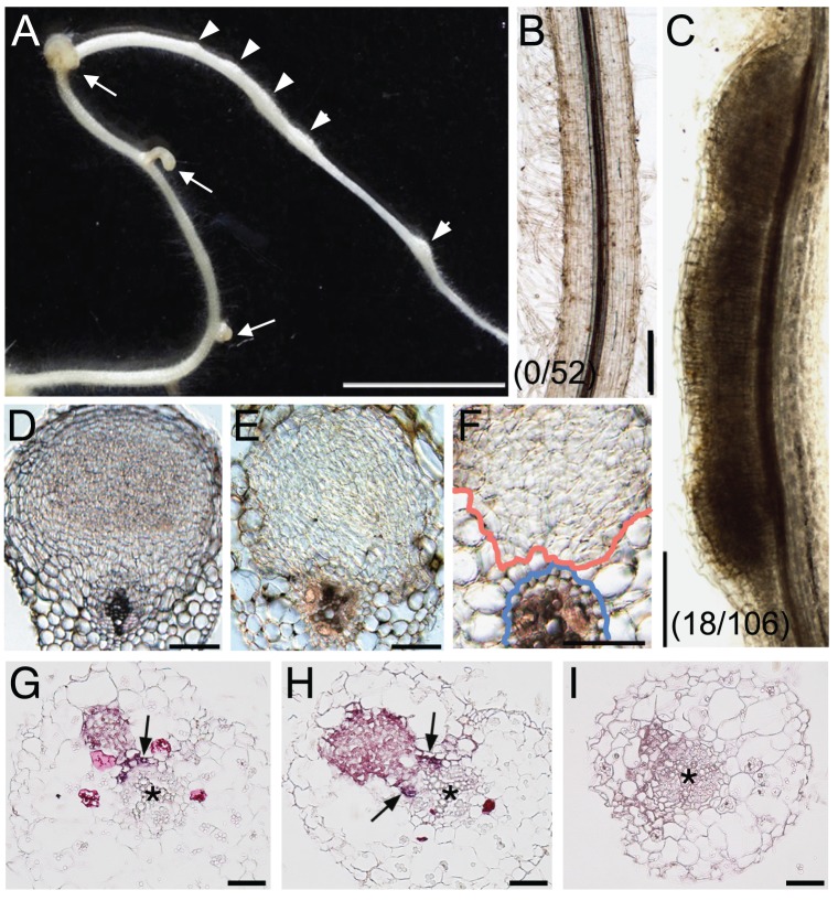
Figure 5. NIN overexpression induces cortical cell division.
(A) A Gifu (wild-type) root was transformed with ProLjUb-NIN and then cultured for 6 weeks in the absence of M. loti. Bumps (arrowheads) and malformed lateral roots (arrows) are indicated. (B,C) Cleared roots that were transformed with either an empty vector (B) or ProLjUb-NIN (C). The fractions of plants with bumps are shown in parentheses. (D) A transverse section of a root nodule primordium (10 dai) formed on a MG-20 (wild-type) root that was transformed with the empty vector. (E,F) Transverse sections of bumps formed on uninoculated MG-20 roots that were transformed with ProLjUb-NIN. Blue and red lines in (F) represent the outer edges of the endodermis and the boundary of the region with dividing cortical cells, respectively. (G–I) in situ RNA hybridization of ENOD40-1 in transverse sections of bumps caused by NIN overexpression, using either antisense (G,H) or sense probes (I). Asterisks indicate the central xylem. Arrows indicate the pericycle with in situ signals. Bars: 5 mm in (A); 0.2 mm in (B,C); 0.1 mm in (D–F); 50 µm in (G–I).

