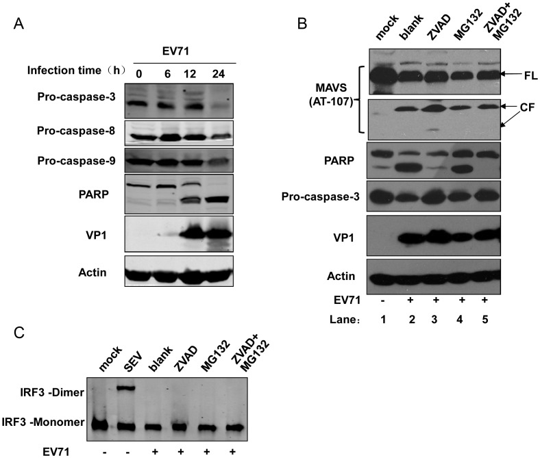
Figure 4. MAVS cleavage is independent of cellular apoptosis and proteasome degradation.
(A) Time course evaluation of caspase 3, 8, and 9 activation and PARP cleavage in EV71-infected HeLa cells (MOI = 10). (B) Effects of caspase and proteasome inhibitors on EV71 induced MAVS cleavage. HeLa cells were mock infected (lane 1) or infected with EV71 (MOI = 10) in the absence (blank, lane 2) or presence of Z-VAD-FMK (ZVAD) (100 µM, lane 3), MG132 (20 µM, lane 4), or both (lane 5). At the 24 h post-infection time point, the cells were harvested and western blot was used to detect MAVS and its cleavage fragments using the anti-MAVS AT107 antibody. (C) Effects of caspase and proteasome inhibitors on IRF3 dimerization. The inhibitor concentrations used in this experiment were the same as in (B).

