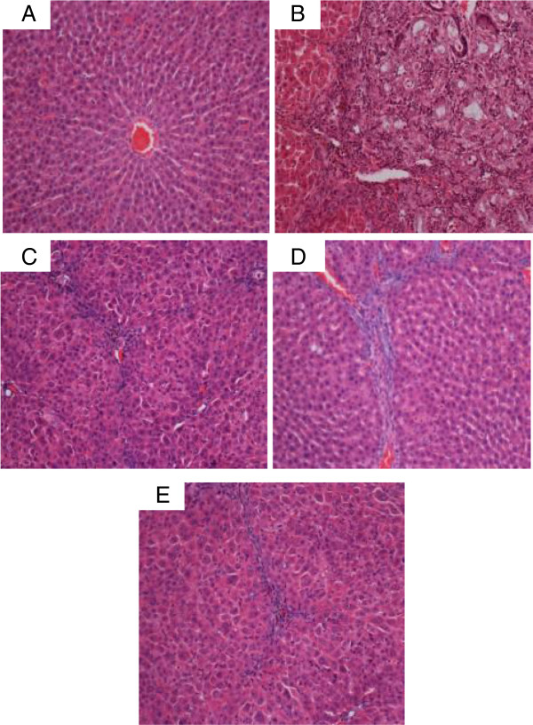
Figure 13.
Examples of representative histopathological sections from livers sampled from rats in different experimental groups. (13A) Normal histological structure and architecture were seen in livers from the control rats. (13B) Severe structural damage, formation of pseudolobules with thick fibrotic septa and necrotic areas were present in the livers of hepatotoxic rats. (13C) Mild inflammation but no fibrotic septae were observed in the liver of the hepatoprotective rat treated with Silymarin. (13D) Partially preserved hepatocytes and architecture with small area of necrosis and fibrotic septa were observed in the livers of rats treated with low dose CLRE. (13E) Partially preserved hepatocytes and architecture with small areas of mild necrosis were observed in the liver of rats treated with high dose CLRE. (H&E stain, original magnification ×20).

