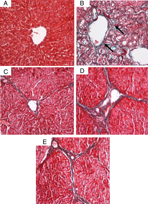
Figure 14.
Masson’s Trichrome staining of representative livers sampled from rats in different experimental groups. (14A) Normal liver structure without signs of collagen deposition in livers from control rats. (14B) Severe collagen deposition (arrow) and severe fibrosis were seen in the livers from cirrhosis rats. (14C) Minimal collagen deposition in the liver of the hepatoprotective rat treated with Silymarin. (14D) Moderated collagen deposition and moderate congestion around the central vein in the livers of rats treated with low dose CLRE. (14E) Mild collagen deposition was observed in the livers of rats treated with high dose CLRE. (Original magnification ×20).

