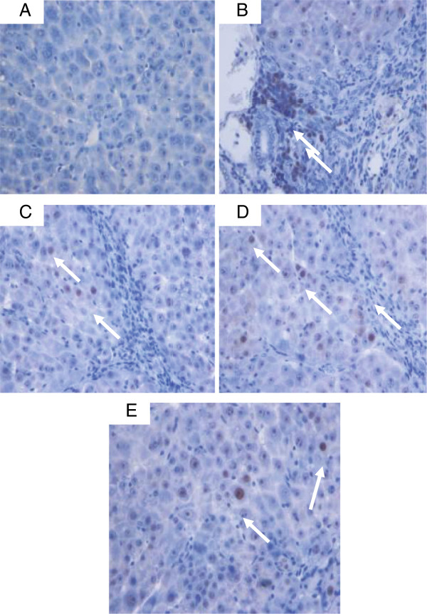
Figure 16.
PCNA staining of livers sampled from rats in different experimental groups. (16A) Normal livers without signs of PCNA expression in hepatocytes from control rats. (16B) Severe fibrosis with greater PCNA expression in the hepatocytes from cirrhosis rats. (16C) Less PCNA-stained hepatocytes (arrow) indicating less hepatocyte regeneration in the liver of hepatoprotected rats treated with Silymarin. (16D) Moderate hepatocyte regeneration as indicated by moderate PCNA staining (arrow) in the liver of rats treated with low dose CLRE. (16E) Minor PCNA expression (arrow) with few regenerative hepatocytes was observed in the liver of rats treated with high dose CLRE. (Original magnification ×40).

