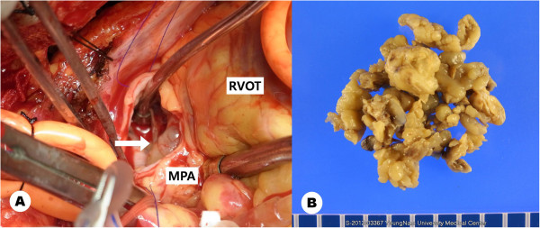
Figure 3.

The large multiple lobulated mass is seen through the main pulmonary artery (arrow). The mass extended distally to both pulmonary artery branches with a retrograde extension to the pulmonary valve and right ventricular outflow tract (left). Resected tumor masses (right). RV: right ventricle, MPA: main pulmonary artery.
