
Figure 7. CaMKII inhibition suppresses SCaW in mdx ventricular myocytes at 3-Hz.
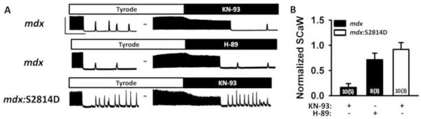
(A) Representative tracings showing spontaneous Ca2+ waves (SCaW) following 3-Hz pacing. CaMKII inhibitor KN-93 (1 μM) or PKA inhibitor H-89 (1 μM) were added subsequently, followed by another observation period for SCaWs. Scale bars: 2 F/F0 (vertical) and 20 seconds (horizontal). (B) Bar graph showing SCaW frequency after inhibitor administration normalized to SCaW frequency in Tyrode.