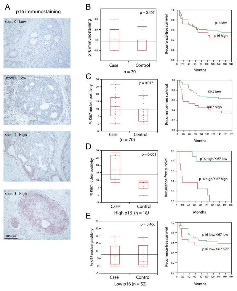
Figure 1. p16 overexpression coupled with proliferation increases the risk of subsequent tumor events among women with DCIS.
A) Representative p16 immunohistochemistry.
B) High p16 staining fails to stratify women with DCIS that develop subsequent disease. Recurrence-free survival plots demonstrate that women with DCIS staining high or low for p16 develop subsequent disease at the same rate.
C) Ki67 index labelling stratifies recurrence-free survival in women with DCIS.
D) DCIS exhibiting high p16 immunostaining and elevated Ki67 identifies women that have a reduced recurrence-free survival.
E) Ki67 does not differentiate risk in DCIS lesions with low p16. Box plots and corresponding p-values were determined using Wilcoxon/Kruskal-Wallis rank of sums test. Survival plots were generated using Kaplan-Meyer analysis.

