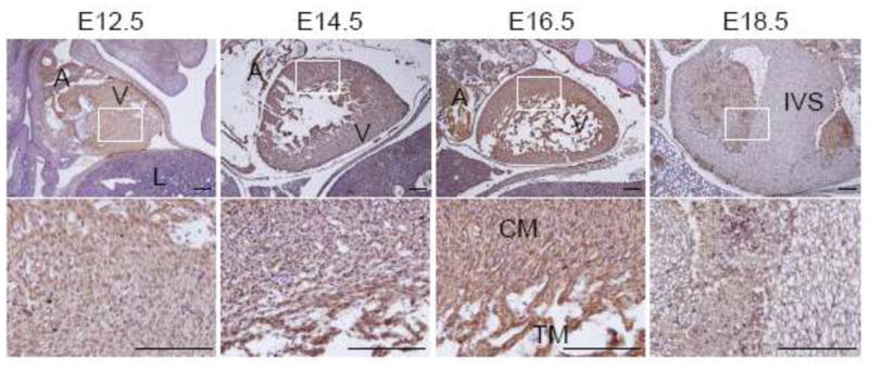
Fig. 4.

Expression of MDA-9/syntenin in the developing heart. Immunohistochemistry was performed at each section at E12.5 to 18.5 of the developing heart. Expression of MDA-9/syntenin at each stage is shown. Every enlarged image in the lower panel corresponds to the boxed area in the upper panel. Scale bars indicate 100 μm. A: atrium, V: ventricle, L: lung, CM: compact myocardium, TM: trabeculated myocardium and IVS: interventricular septum.
