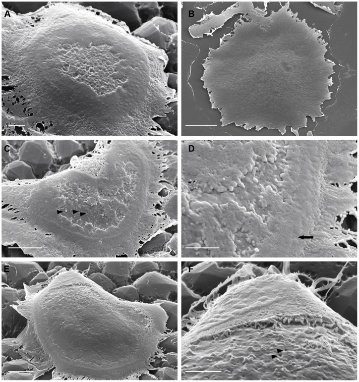
Figure 1. The apical surface of osteoclasts incubated on vitronectin- or fibronectin-coated nail-varnish.
Osteoclasts were incubated on coverslips coated with nail-varnish and then vitronectin (A, C–F) or fibronectin (B), fixed, and inverted onto glass slides before removal of nail varnish and preparation for SEM. A: cell incubated on vitronectin, showing a circular arrangement of nodules, which merge in places (at top left of the cell) into ridges. These surround a central region filled with membrane folds. B: substrate-apposed surface of cell incubated on fibronectin lacks these features. C, D: cell shows raised nodules that correspond to podosomes (arrow). Within this podosome crescent are irregular, predominantly peripheral, patches of ruffled membrane. Orifices can be seen in the fold-free surface (arrowheads). E, F: well-spread cell with podosomes predominantly merged into ridges. Folded membrane is flattened against the substrate and is limited to a peripheral strip. Orifices can be seen in the apical membrane central to the peripheral ruffles (F, arrowheads). D and F are magnified portions of C and E, respectively. Bar 2 μm ( A, D, F), 5 μm (C, E) and 10 μm (B).

