
Figure 2. Transmission electron micrographs of osteoclast after incubation on vitronectin-coated tissue culture plastic.
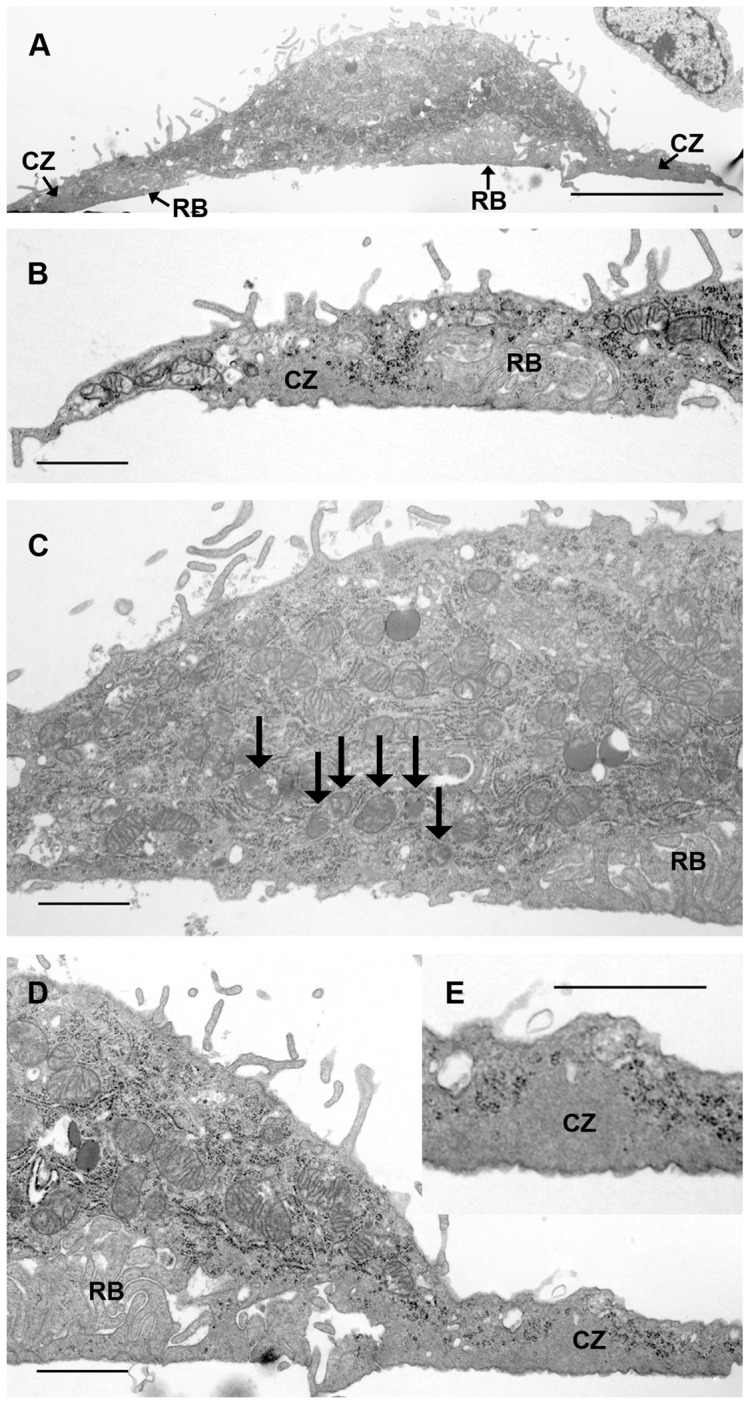
A: low magnification of cell shown in B–E. In B, D a peripheral ‘clear zone’ (CZ), clear of organelles, is seen. This is further magnified in D and E, where it is immediately peripheral to a zone of closely-packed membrane folds (ruffled border, RB), which show a pale appearance resembling that of ‘clear zones’. C: central area is devoid of membrane folds. Many vesicles containing electron-dense material (arrows) can also be seen above this area. Bar 5 μm (A) and 1 μm (B–E).