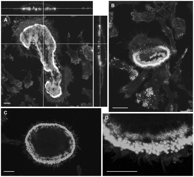
Figure 4. CLSM images of F-actin in osteoclasts after incubation on bone or vitronectin-coated glass coverslips.
A: Osteoclast incubated on bone shows strong staining for F-actin in actin ring, but also significant F-actin staining within the ring. The latter is patchy with intervening F-actin-free regions. B: an osteoclast incubated on vitronectin-coated glass coverslip shows a crescent of intensely-F-actin-positive podosomes. F-actin is also present adjacent to the podosome crescent, and as more central patches. C, D: patches of F-actin immediately inside the strongly-positive podosome ring of osteoclast on vitronectin. Bar 10 µm.

