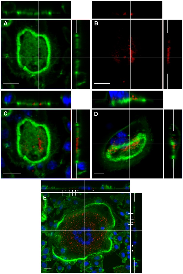
Figure 6. CLSM localization of F-actin (green) and cathepsin K (red) in osteoclasts.
A–D: Osteoclasts incubated on bone show patchy distribution of F-actin within the F-actin ring (clearly shown in panel A). In the cell with a non-migratory appearance (A–C), cathepsin K localizes towards the centre of the apex (B), in a region that is free of F-actin (A, C). In the osteoclast showing a migratory morphology (D), cathepsin K localizes to the retracting pole, also in an F-actin-free region (D, z-stacks). E: In osteoclast incubated on vitronectin, cathepsin K localizes to punctuate foci present at the cell-substrate interface (arrows) centrally to F-actin ring. C-E are counterstained with DAPI. Bar 10 µm.

