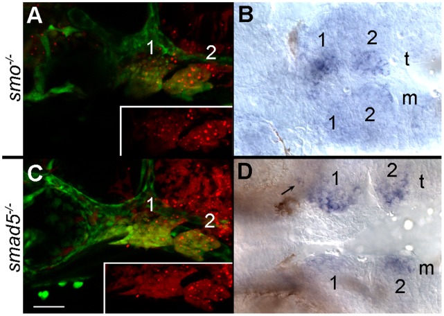
Figure 2. Neural crest cells require the reception of Bmp and Hh signaling to express satb2.
(A, C) Lateral views at 36 hpf; (B, D) ventral views at 36 hpf. Both donor and host embryos were fli1:EFGP transgenics to allow for visualization of neural crest cells. (A) Wild-type crest transplanted into the arches of smo mutant embryos are shown in red in the inset. (B) smo+ crest restores the ventral arch expression of satb2 on the side of the transplant (t) as compared to the mutant side of the embryo (m). (C) Lateral views of a 36 hpf smad5 mutant that received a transplant of smad5+ neural crest. The distribution of smad5+ cells (red in inset) is shown. (D) Expression of satb2 is restored in the ventral pharyngeal arch and in the palatal precursors (arrow) on the transplanted side of the embryo. Arches 1 & 2 are numbered in each panel.

