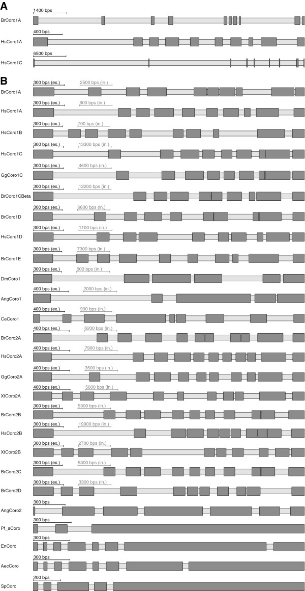
Figure 1.
Gene structure schemes of coronins. A) The schemes illustrate examples of coronin genes from human (Hs = Homo sapiens) and zebrafish (Br = Brachydanio rerio) Dark grey bars and light grey bars mark exons and introns, respectively. B) This figure illustrates examples of coronin genes from vertebrates (Hs = Homo sapiens, Br = Brachydanio rerio, Gg = Gallus gallus, Xt = Xenopus tropicalis), arthropods (Dm = Drosophila melanogaster, Ang = Anopheles gambiae), nematods (Ce = Caenorhabditis elegans) and the protozoan parasite Plasmodium falciparum (Pf_a). In order not to make small exons vanish when very large intronic stretches are present, the scaling of introns and exons is automatically balanced to make the picture visually meaningful (scale bars for exons and introns are given, respectively). Here, except for AngCoro2 and Pf_aCoro in all schemes the introns were scaled down and the exons scaled up so that the average length of the introns equals the average length of the exons.

