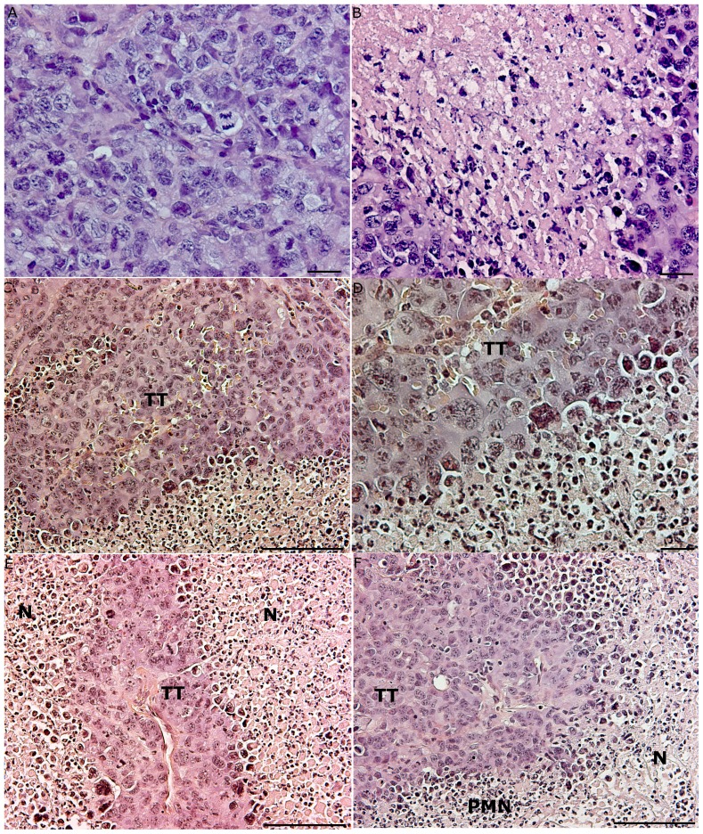
Figure 4. Histology of tumor tissues from mice with or without AT.
(A) Fourteen days after inoculation, the tumor tissue was dense with mitotic figures, and no infiltration of immune cells was observed (HE stain, bar 20 µm). (B) A section of tumor tissue (TT) that reached the plateau phase of growth 22 days after inoculation. A large area of necrosis (N) was visible, but no infiltrating immune cells were present (HE stain, bar 20 µm). (C) Massive infiltration of PMNs in S180 tumors after AT of cancer-resistant SR/CR leukocytes to cancer susceptible C57BL/6 mice (representative sections, n = 8). Tumor tissue (TT) from a mouse with tumor regrowth showed large areas of infiltrating PMNs (PMN) and necrosis (N) (HE stain, bar 100 µm). (D) Tumor tissue (TT) from a mouse with tumor regrowth revealed the infiltration of PMNs (PMN) (HE stain, bar 20 µm). (E) The regression of tumor tissue (TT) in a cancer-susceptible C57BL/6 mouse after AT of SR/CR leukocytes. Large necrotic areas (N) in the tumor tissue were observed (HE stain, bar 100 µm). (F) An image of the same tumor as in C), showing a clear demarcation between the vital tumor tissue (TT) and the infiltrating PMNs (PMN), as well as large necrotic (N) areas (HE stain, bar 100 µm).

