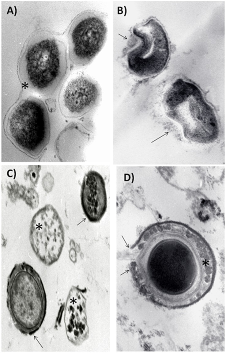
Figure 3. Representative electron microscopy micrographs of Mycobacterium tuberculosis strain H37Rv treated with high concentrations of IDR peptides.
(A) Control bacteria show thick homogeneous electron lucent cell wall (asterisk) and cytoplasm with some electron dense granules (63,000x); (B) Incubation with peptide 1002 induces abnormalities in the cell wall, such as thinning, budding and partial dissolution (arrows) (100,000x). (C) Peptide HH2 induces complete disappearance of the cell wall with electron lucent cytoplasm (asterisks) or homogenous cell wall thinning and condensation (arrows) with slight electron lucent change of the cytoplasm (80,000x). (D) Incubation with peptide 1018 produced a peripheral electron dense rim with areas of rupture and dissolution (arrows) in the cell wall, which also have irregular electron dense condensations (asterisk) and an electron lucent halo around the cytoplasm (100,000x). All bars in the figures represent 5 µm. The micrographs are representative of 2 independent experiments.

