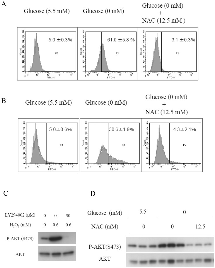
Figure 2. ROS mediates AKT phosphorylation under glucose deprivation.
(A)(B)(D) HepG2 cells were cultured in either glucose-containing medium or glucose-deprived medium in the absence or presence of 12.5 mM of NAC for 0.5 h. ROS production was measured using flow cytometry. Cells were stained with (A) 5 µM of DCFDA or (B) 5 µM of BES-H2O2. Cells were gated within a range contained in the upper 5% of the total cell count under the glucose replete condition. (D) The AKT phosphorylation level was evaluated by immunoblotting. (C) Addition of H2O2 to media containing 5.5 mM of glucose in the absence or presence of 30 µM of LY294002 for 0.5 h, followed by immunoblotting.

