Figure 3.

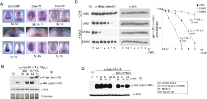
Antagonism between Smurf1 and Smurf2 in the regulation of ephrinB1 degradation. (A) Whole-mount in situ hybridizations of Smurf1, Smurf2, and ephrinB1 in stage 16–19 embryos (for top and bottom panels, left photo is anterior view, and right photo is dorsal view; middle panels represent cross sections of stage 16–17 embryos, and the level of section is represented by a thin horizontal line in the top right panels). (B) Western blot analysis of dorsal axial tissue from stage 16 embryos show that introduction of Smurf1 MO reduces exogenously expressed HA-tagged ephrinB1, while Smurf2 MO increases ephrinB1 in this tissue. Blots were also probed with anti-Erk antibodies and Ponceau-stained as loading controls. (C) Western blot analysis of dorsal axial tissue from stage 14 embryos previously injected with HA-tagged ephrinB1 RNA that was titrated to yield roughly equivalent ephrinB1 protein levels in the presence of the indicated MOs. The explanted tissue was treated with cycloheximide (CHX) for 5 h, and Western analysis shows that introduction of Smurf1 MO reduces exogenously expressed ephrinB1 by half within 60 min, while Smurf2 MO increases ephrinB1 half-life to ∼5 h in this tissue. Blots were also probed with anti-Erk antibodies as loading controls. The quantification of the data from three independent experiments is displayed in the right panel; error bars show SD. (D) One- or two-cell stage embryos were left uninjected (UI) or injected with HA-tagged ephrinB1 (100 pg) RNA alone or along with Smurf1 MO (10 ng). At stages 16–18, dorsal axial tissue was dissected and treated with DMSO carrier (D), chloroquine (C), MG132 (M), or dynasore (DS) at the indicated concentrations for 4–5 h. Samples were analyzed directly by SDS-PAGE, then immunoblotted with anti-HA antibodies or anti-Erk antibodies to reveal exogenous expression levels of ephrinB1 and the endogenous Erk levels (as a loading control), respectively.
