FIGURE 1.
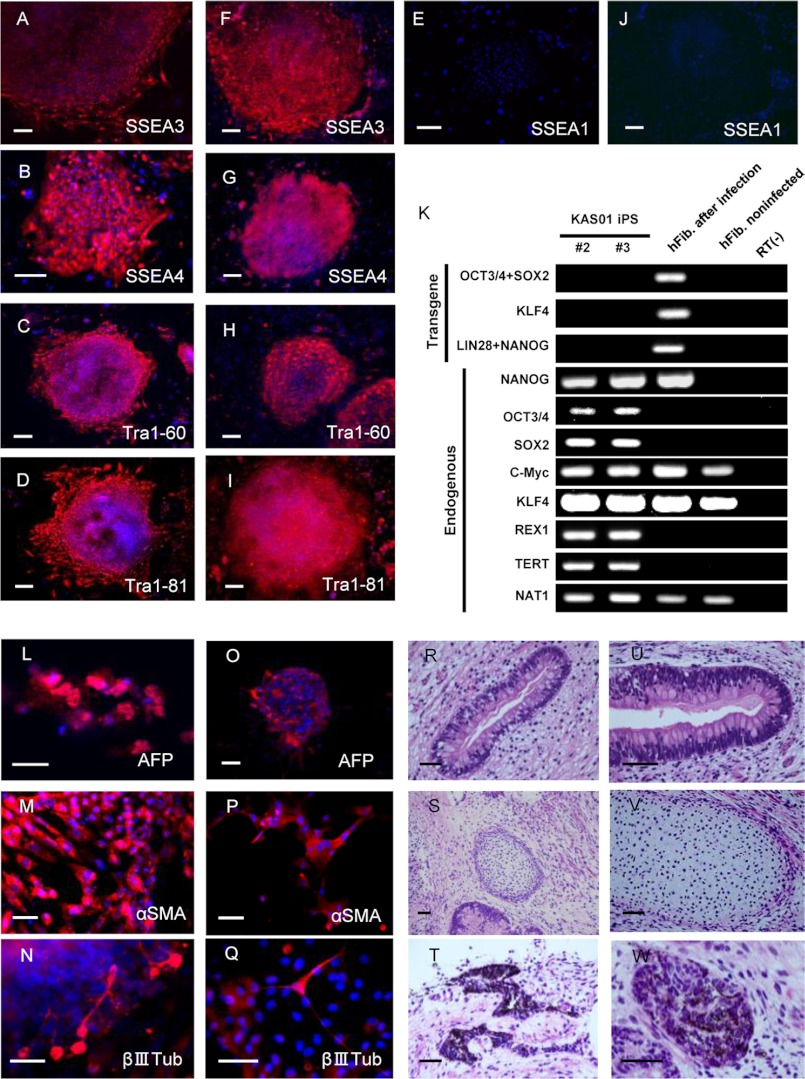
Generation of KAS01 #2 and #3 iPSCs and determination of their pluripotency. A–J, both KAS01 #2 and #3 iPSC lines exhibit markers of pluripotency (A–E, KAS01 #2; F–J, KAS01 #3). All iPSCs expressed the pluripotency markers Tra-1–60, Tra-1–81, SSEA3, and SSEA4. SSEA1 is used as a negative surface marker for undifferentiated stem cells. The nuclei were stained with DAPI. Scale bar, 200 μm. K, RT-PCR analysis of the transgenes OCT3/4, SOX2, KLF4, and the endogenous human embryonic stem cell marker genes. Patient fibroblasts 6 days after retroviral transduction are positive for the transgenes. hFib, healthy control fibroblast; RT(−), healthy control samples without RT-PCR. L–Q, embryoid bodies derived from KAS01 #2 and KAS01 #3 iPSCs express germ layer-specific markers, including α-fetoprotein (AFP, endoderm), α-smooth muscle actin (αSMA, mesoderm), and βIII-tubulin (βIIITub, ectoderm). L–N, KAS01 #2, O–Q: KAS01 #3. Scale bar, 100 μm. R–W, teratomas derived from SCID mice injected with KAS01 #2 and KAS01 #3 iPSCs. Representative images of hematoxylin and eosin staining of teratoma sections are shown (R–T, KAS01 #2; U–W, KAS01 #3). Tissues representing all three embryonic germ layers, including glandular structure (endoderm), cartilage (mesoderm), and pigmented epithelium (ectoderm), are visible in cells of both iPSC lines. Scale bar, 50 μm.

