FIGURE 5.
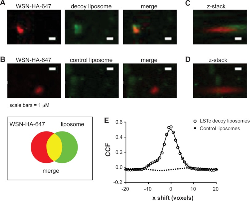
LSTc decoy liposomes bind directly to influenza A virus. Immunofluorescent Z-stack images were obtained of WSN HA-647 and fluorescent liposomes, viewed from above (A and B) or rotated 37 degrees (C and D). A and C, LSTc decoy liposomes, which contain NBD (green) bound specifically to WSN HA-647 (red) and can be seen binding to the apical portion of virus foci when the field of view is rotated 37 degrees. B and D, control liposomes do not show significant co-localization with WSN HA-647. Scale bar, 1 μm. E, Van Steensel's CCF coefficient analysis of images A and B is shown. The CCF is the Pearson coefficient of WSN HA-647 and liposomes at each pixel shift in the x dimension; this calculation is based on a representative field of 45 by 45 μm. Decoy liposomes (open circles) significantly correlate to WSN HA-647 (p < 0.001), whereas control liposomes (black squares) do not (p = 0.21).

