FIGURE 1.
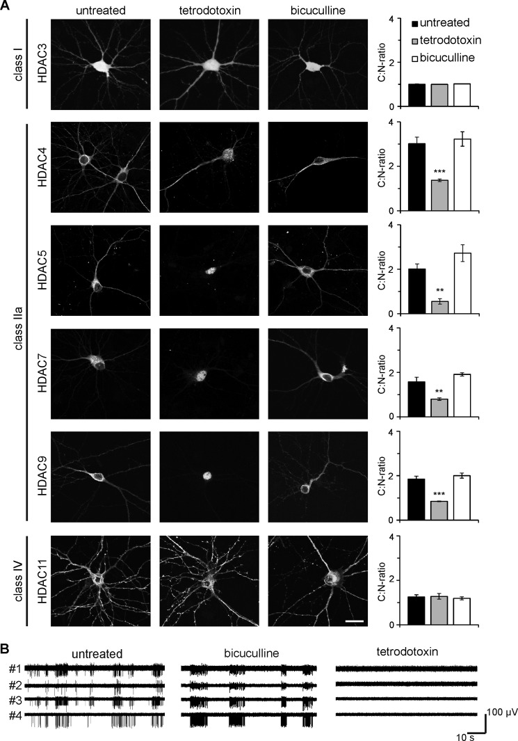
Nucleo-cytoplasmic shuttling of class IIa HDACs is dependent on neuronal activity. A, representative micrographs of cultured hippocampal neurons transfected with epitope-tagged HDAC constructs (HDAC3-HA, HDAC4-FLAG, HDAC5-FLAG, HDAC7-HA, HDAC9-HA, and HDAC11-HA) and either left untreated or treated with TTX or bicuculline as indicated. Scale bar is 20 μm. Graphs show the ratio between cytoplasmic and nuclear localization. 400–1000 cells were analyzed for each tested HDAC and experimental condition from a minimum of three independent preparations. Statistically significant differences are indicated with asterisks (***, p < 0.001; **, p < 0.01, one-way ANOVA, Dunnett's post hoc test). Error bars, S.E. B, typical examples of MEA recordings obtained from untreated cultured hippocampal neurons (left), and neurons after exposure to bicuculline (50 μm, center), or TTX (1 μm, right). Simultaneous recordings from four separate electrodes are shown in each panel.

