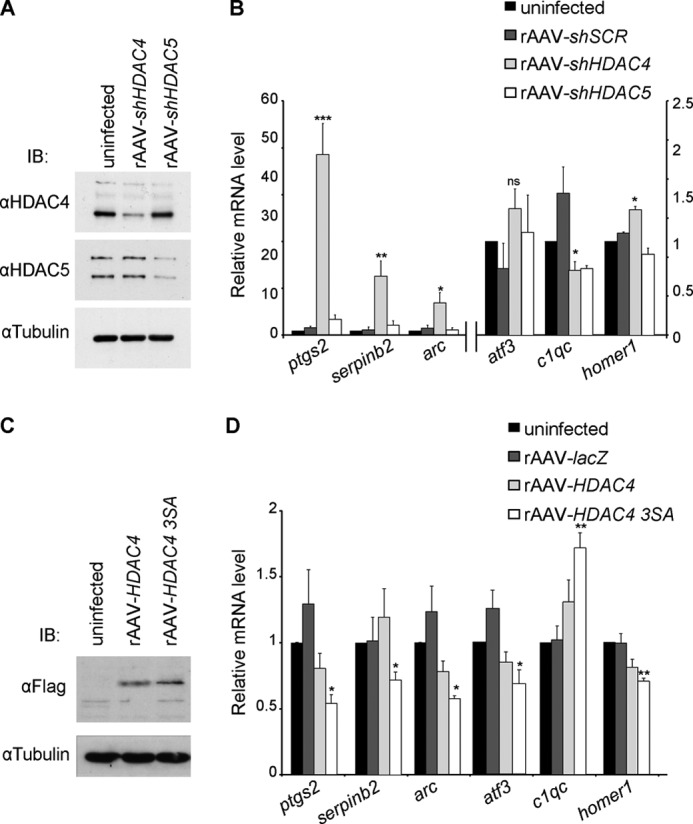
FIGURE 7.

HDAC4 regulates expression of genes involved in neuroadaptations. A, immunoblot analysis of uninfected hippocampal neurons and of hippocampal neurons infected with the indicated rAAVs. Tubulin immunoblot is shown as control for protein loading. B, QRT-PCR analysis of expression of ptgs2, serpinb2, arc, atf3, c1qc, and homer1 in uninfected hippocampal neurons and in hippocampal neurons infected with rAAV-shSCR, rAAV-shHDAC4, or with rAAV-shHDAC5 (n = 5). ptgs2, arc, and serpinb2 values refer to the axis on the left. C, immunoblot analysis of uninfected hippocampal neurons and of hippocampal neurons infected with the indicated rAAVs. rAAV-HDAC4 and rAAV-HDAC4 3SA all carry a FLAG cassette which was used for detection via an anti-FLAG antibody. Tubulin immunoblot is shown as control for protein loading. D, QRT-PCR analysis of ptgs2, serpinb2, arc, atf3, c1qc, and homer1 expression in uninfected hippocampal neurons and in hippocampal neurons infected with rAAVs giving rise to the indicated proteins (n = 5). Statistically significant differences are indicated with asterisks (***, p < 0.0005; **, p < 0.05; *, p < 0.05 one-way ANOVA, Dunnett's post hoc test). Error bars, S.E.
