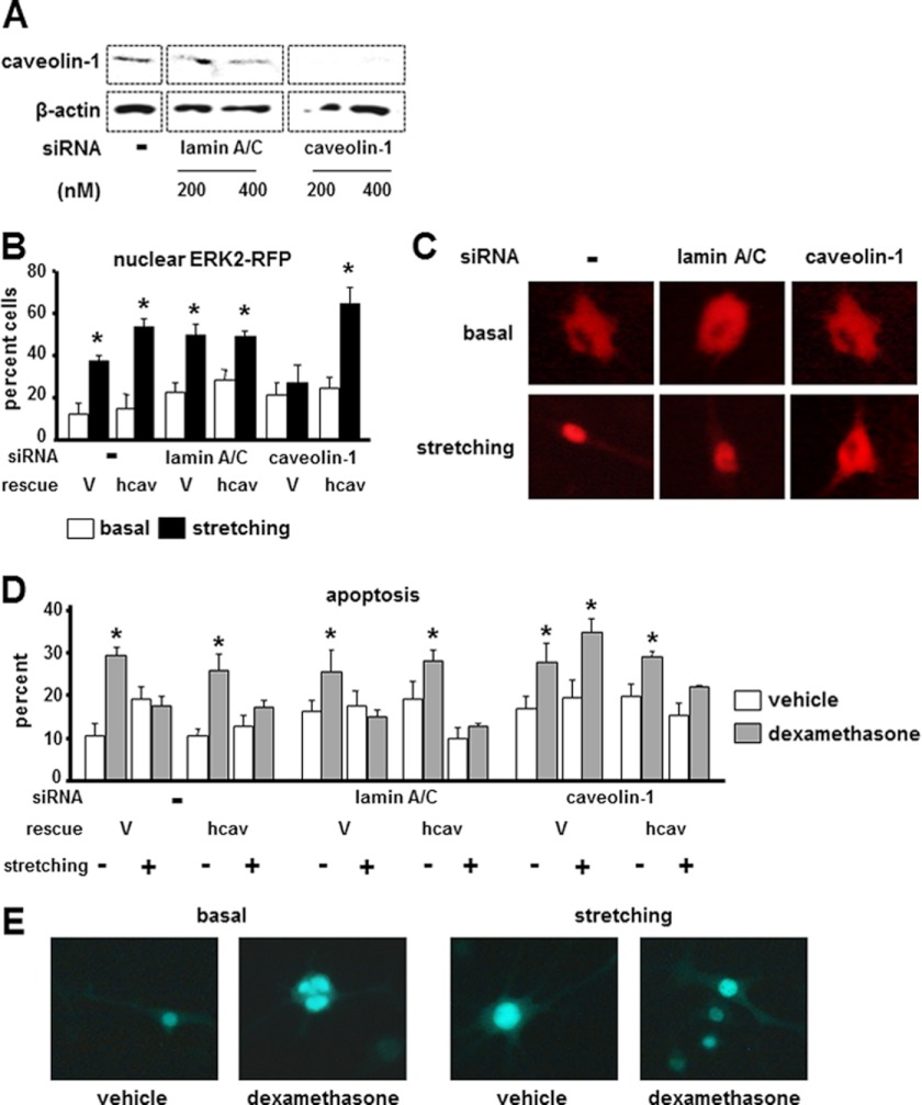
FIGURE 1.
ERK nuclear translocation and anti-apoptosis induced by stretching are abolished by knocking down caveolin-1. A, caveolin-1 protein expression was determined by Western blot analysis in MLO-Y4 cells treated 200 or 400 nm siRNA oligonucleotides. β-Actin levels show equal loading. B–E, caveolin-1 expression was silenced in MLO-Y4 cells using 200 nm siRNA. Additional cultures were left untreated (−) or were silenced for lamin A/C (as controls), followed by transfection with empty vector (V) or human caveolin-1 (hcav) constructs together with ERK2-RFP and MEK to allow quantification of ERK nuclear translocation and with nGFP to allow quantification of apoptosis. 24 h later, cells were mechanically stimulated by stretching at 5% elongation for 10 min, and ERK nuclear translocation (B and C) and apoptosis (D and E) were quantified as described under “Experimental Procedures.” Error bars indicate means ± S.D. of triplicate determinations. *, p < 0.05 versus basal or vehicle for each construct in B and D, respectively, by one-way ANOVA. Representative images are shown exemplifying cells with cytoplasmic and nuclear ERK2-RFP (C) or live and apoptotic nGFP-expressing cells (E).

