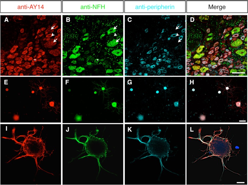
FIGURE 4.
Immunofluorescence localization of Nes-S protein in DRG neurons of adult rats. A–D, triple labeling of DRG tissue sections of adult rats with anti-AY14 (A), anti-NFH (B), and anti-peripherin (C). All three populations of the DRG neurons, including NFH+/peripherin− large neurons (arrows), NFH+/peripherin+ medium neurons (asterisks), and NFH−/peripherin+ small neurons (arrowheads), were AY14+. D shows merged images. Scale bar: 50 μm. E–H, triple labeling of primary DRG neurons of adult rats with anti-AY14 (E), anti-NFH (F), and anti-peripherin (G). Anti-AY14-IR was observed in all of the neurons. H shows merged images. Scale bar: 50 μm. I–L, triple labeling of primary DRG neurons of adult rats with anti-AY14 (I), anti-NFH (J), and anti-peripherin (K) by high magnification confocal microscopy. The IFs formed a cage-like cytostructure at the periphery of neurons and extended into the neurites. L shows merged images. Scale bar: 15 μm.

