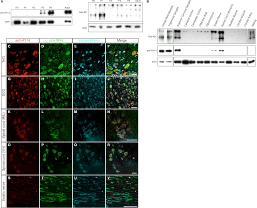
FIGURE 6.
Postnatal expression and tissue distribution of Nes-S. A, immunoblotting of IF-enriched preparations of DRG from rats of different postnatal days, as well as the adult rats, with anti-AY14 and Rat-401. Nes-S expression was not detected until P5, and its intensity reached the highest point at adult. B, immunoblotting of IF-enriched preparations of various tissues of adult rats with anti-AY14 (left) and Rat-401 (right). Nes-S was expressed in DRG, TriG, SCG, and thoracic spinal cord, whereas a smaller amount of Nes-S was detected in sciatic nerve. C–V, triple labeling of TriG, SCG, spinal cord, and sciatic nerve of adult rats with anti-AY14, anti-NFH, and anti-peripherin. In TriG (C–F) and SCG (G–J), all neurons were AY14+. In spinal cord, the preganglionic sympathetic neurons (K–N), and motor neurons (O–R) were AY14+. In sciatic nerve, a trace amount of anti-AY14-IR was detected in the axoplasm of neurites (S–V). IML, intermediate lateral horn; VH, ventral horn. Scale bar: 50 μm.

