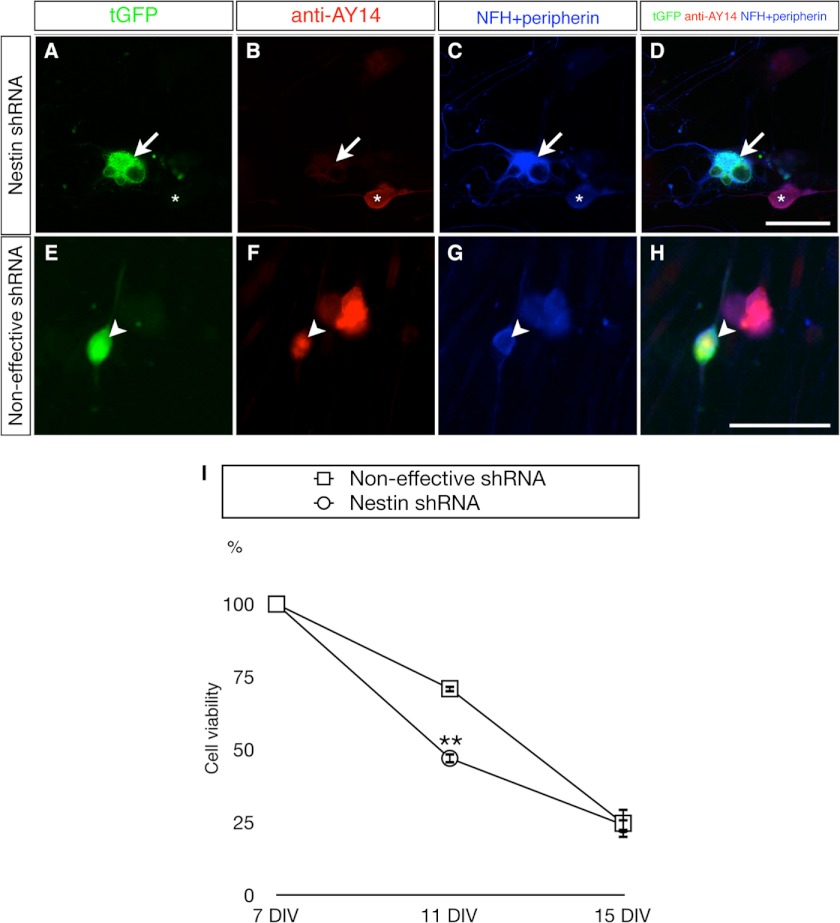
FIGURE 8.
Impaired cell survival in primary DRG neurons of adult rats upon RNAi knockdown of Nes-S. A–D, reduced expression of Nes-S by nestin shRNA expression vector. The transfected neurons in the 7 DIV cultures were recognized by their tGFP expression (A) and morphological features, i.e. a rounded soma and elongated neurites. Triple labeling with anti-AY14 (B) and anti-NFH and anti-peripherin (C, their IRs were recorded in the same channel) showed that anti-AY14-IR intensity was reduced in DRG neurons expressing the tGFP reporter gene of shRNA vector (arrow). The anti-AY14-IR remained in the untransfected neurons (asterisk). D shows merged images. E–H, expression level of Nes-S was not altered by noneffective shRNA control vector. The tGFP+ neurons (E, arrowhead) present AY14-IR (F) as well as NFH- and peripherin-IR (G). H shows merged images. Scale bar: 50 μm. I, reduced cell survival rate in Nes-S knockdown DRG neurons during prolonged culture. Live cell images of tGFP+ neurons were taken by an inverted fluorescence microscope at 7, 11, and 15 DIV. At each day, the number of tGFP-expressing neurons was counted. The viability of Nes-S knockdown neurons was significantly decreased at 11 DIV (n = 3, **, p < 0.01, two-tailed t test). Neurons transfected with the noneffective shRNA vector were used as control. The survival rate of each DIV was calculated using the number of tGFP+ cells of the same plate at 7 DIV as the denominator. For the number of neurons of both transfected groups at each day, see supplemental Table S2. Error bars indicate S.E.

