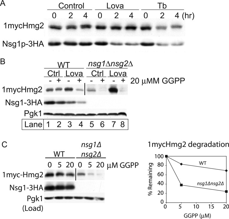
FIGURE 5.
Interplay of sterol and isoprenoid signals on Hmg2 and Nsg1. A, effect of blocking the sterol pathway before or after GGPP synthesis on 1mycHmg2 and Nsg1–3HA protein levels. Cells were treated with lovastatin (Lova) (25 μg/ml) or Tb (10 μg/ml) for 4 h at 30 °C. At the indicated time points, 4 OD equivalents of cells were harvested and lysed in SUME. Following SDS-PAGE, 1mycHmg2 and Nsg1–3HA were detected by immunoblotting. Similar experiments were performed at least four times with similar results. B, GGPP reversed the stabilization of Hmg2 by lovastatin treatment. WT and nsg1Δnsg2Δ cells were treated with vehicle or lovastatin (6.25 μg/ml). After 1 h at 30 °C, each culture was split in half and treated with methanol ammonia (vehicle) or with 20 μm GGPP for 1 h. After lysis and SDS-PAGE, 1mycHmg2, Nsg1–3HA, and Pgk1 (load control) were detected by immunoblotting. Similar experiments were performed at least four times with similar results. C, Hmg2 and Nsg1 protein levels in a GGPP dose-response experiment. WT and nsg1Δnsg2Δ cells were treated with 0, 5, or 20 μm GGPP at 30 °C for 1 h. Samples were harvested, and immunoblotting was performed as in B. In B and C, a darker exposure is displayed for the nsg1Δnsg2Δ strain because Hmg2 levels are so low. The vertical bar marks where the image was cut. In the graph showing the amount of Hmg2 remaining, the value is compared with the 0 sample and normalized to the load control (Pgk1). Similar experiments were performed at least three times.

