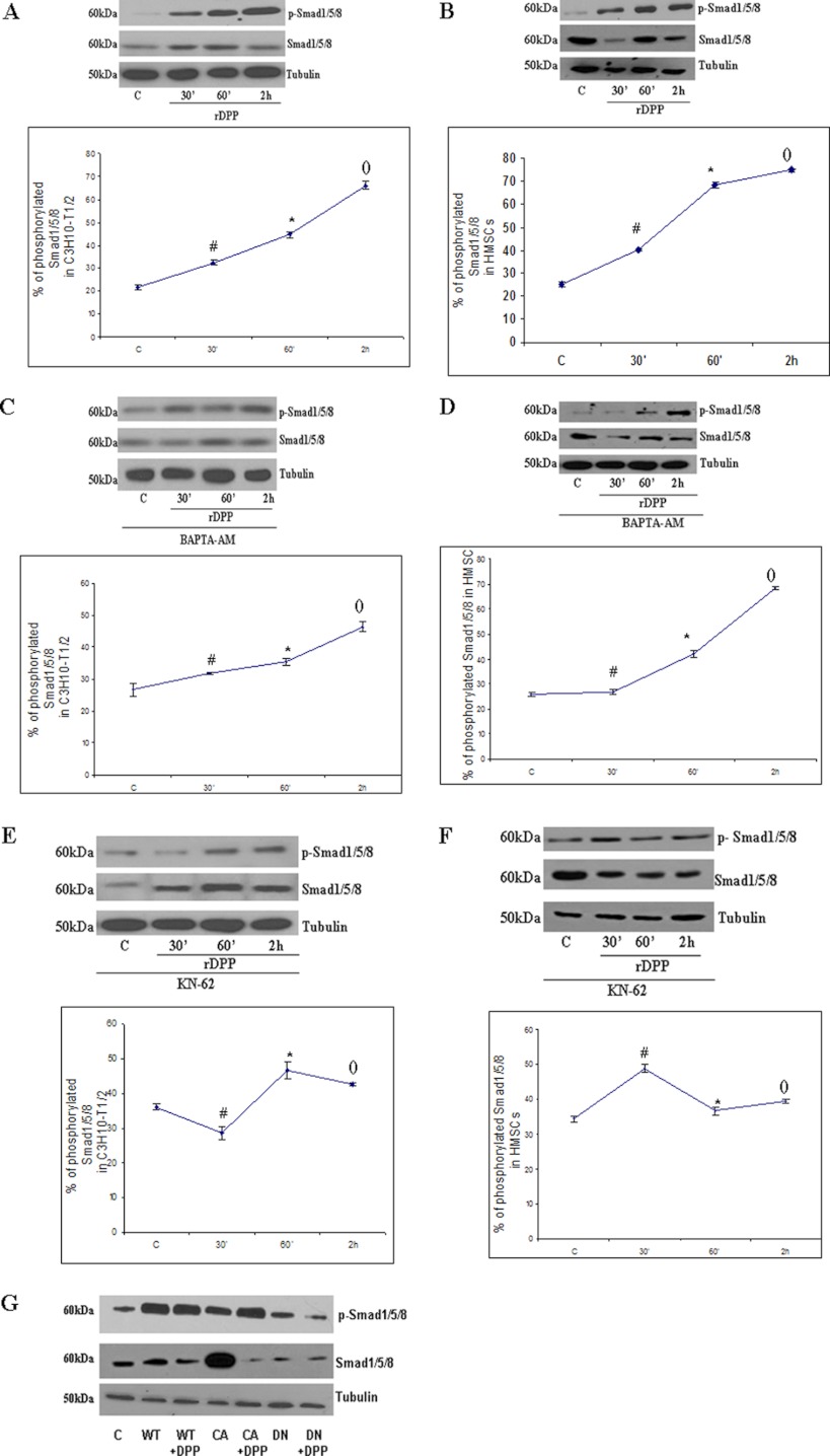
FIGURE 4.
Activation of Smad1/5/8 in C3H10T1/2 and HMSCs by DPP can be inhibited by the treatment with BAPTA-AM and KN-62. A, C3H10T1/2 cells were stimulated with rDPP (500 ng/ml), and Western blotting was performed with Abs against Smad1/5/8. *, #, and (), p < 0.05 as compared with control cells. B, HMSCs were stimulated with rDPP (500 ng/ml), and Western blotting was performed with Abs against Smad1/5/8. *, #, and (), p < 0.05 as compared with control cells. C, C3H10T1/2 cells were pretreated with BAPTA-AM (50 μm) followed by stimulation with rDPP for the indicated time points, and Western blotting was performed with Abs against Smad1/5/8. *, #, and (), p < 0.05 as compared with control cells. D, HMSCs were treated with BAPTA-AM (50 μm) followed by stimulation with rDPP, and Western blotting was performed with Abs against Smad1/5/8. *, #, and (), p < 0.05 as compared with control cells. E, treatment of C3H10T1/2 cells with KN-62 (10 μm) inhibits Smad1/5/8 activation. Total proteins were isolated, and Western blots were developed with anti-Smad1/5/8 antibody and anti-phospho-Smad1/5/8 antibody. Equal loading of the proteins was confirmed by stripping the blot followed by probing it with tubulin. *, #, and (), p < 0.05 as compared with control cells. F, inhibition of Smad1/5/8 in KN-62-treated HMSCs, and Western blotting was performed with Abs against Smad1/5/8. *, #, and (), p < 0.05 as compared with control cells. G, C3H10T1/2 cells were transduced with wild type (WT), constitutively active (CA), and dominant negative (DN) CaMKII and then stimulated with rDPP. Western blotting was performed with Abs against Smad1/5/8. C, control.

