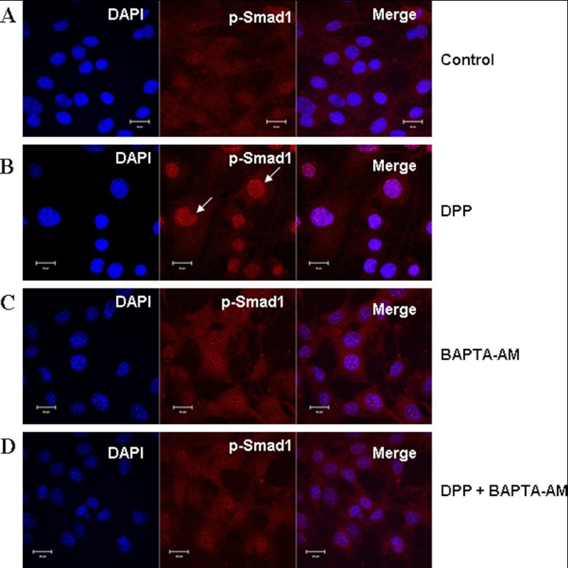
FIGURE 5.
Nuclear localization of p-Smad1 in C3H10T1/2 cells stimulated by DPP and abrogated by BAPTA-AM. C3H10T1/2 cells were stimulated with rDPP (500 ng/ml) or BAPTA-AM (50 μm) for 1 h. The cells were fixed and immunostained for p-Smad1 (red). Control cells exhibited diffused staining throughout the cells for p-Smad1 (A). Upon stimulation with DPP for 1 h, nuclear translocation of p-Smad1 was observed. Arrows indicate nuclear localization of p-Smad1 (B). Cells treated with BAPTA-AM alone displayed cytoplasmic staining of p-Smad1 (C). However, DPP-stimulated and BAPTA-AM-pretreated cells failed to show nuclear translocation of p-Smad1 (D). DAPI (blue) was used to stain the nucleus. Bar, 10 μm.

