FIGURE 8.

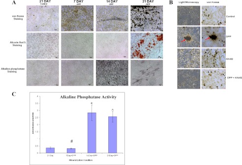
DPP stimulates mineralized nodule formation. C3H10T1/2 cells were cultured in mineralization media for 30 days either with rDPP (500 ng/ml) or KN-62 (10 μm) or both. von Kossa, Alizarin Red S, and alkaline phosphatase staining were performed (A). Terminal differentiation and mineralized nodule formation were observed when cells were stimulated with rDPP. However, untreated control cells, cells treated with inhibitor alone, and cells treated with the inhibitor in the presence of DPP did not show evidence of mineralized nodule formation (B). Arrows point to the mineralized nodule formed. Alkaline phosphatase activity was highly expressed at 14 and 21 days in DPP-treated cells (C). *, #, and [caret], p < 0.05 as compared with control cells. Bar, 20 μm.
