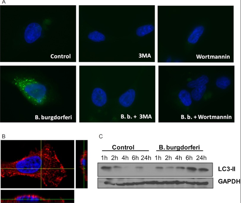
FIGURE 1.
Activation of autophagy by B. burgdorferi. A, GFP-LC3-expressing HeLa cells were preincubated for 1 h at 37 °C in RPMI 1640 medium alone, containing 10 mm 3MA or 100 nm wortmannin in which inhibitors of lysosomal fusion such as ammonium chloride (20 mm) and leupeptine (100 mm) have been added. After 2 h of stimulation with culture medium or B. burgdorferi (m.o.i., 0.4), cells were fixed, nuclei stained with DAPI (blue), and slides were analyzed by fluorescence microscopy. Data are representative of at least four experiments. B, HeLa cells were exposed to GFP-labeled B. burgdorferi (m.o.i., 1) for 2 h, cells were fixed, membrane stained with anti HLA class I Alexa Fluor 649 (red), and nuclei stained with DAPI (blue). Slides were analyzed by confocal microscopy. Data are representative of at least four experiments. C, Western blot analysis of LC3-II in lysates of mouse BMDMs stimulated with B. burgdorferi (m.o.i., 5) was performed for the indicated periods of time.

