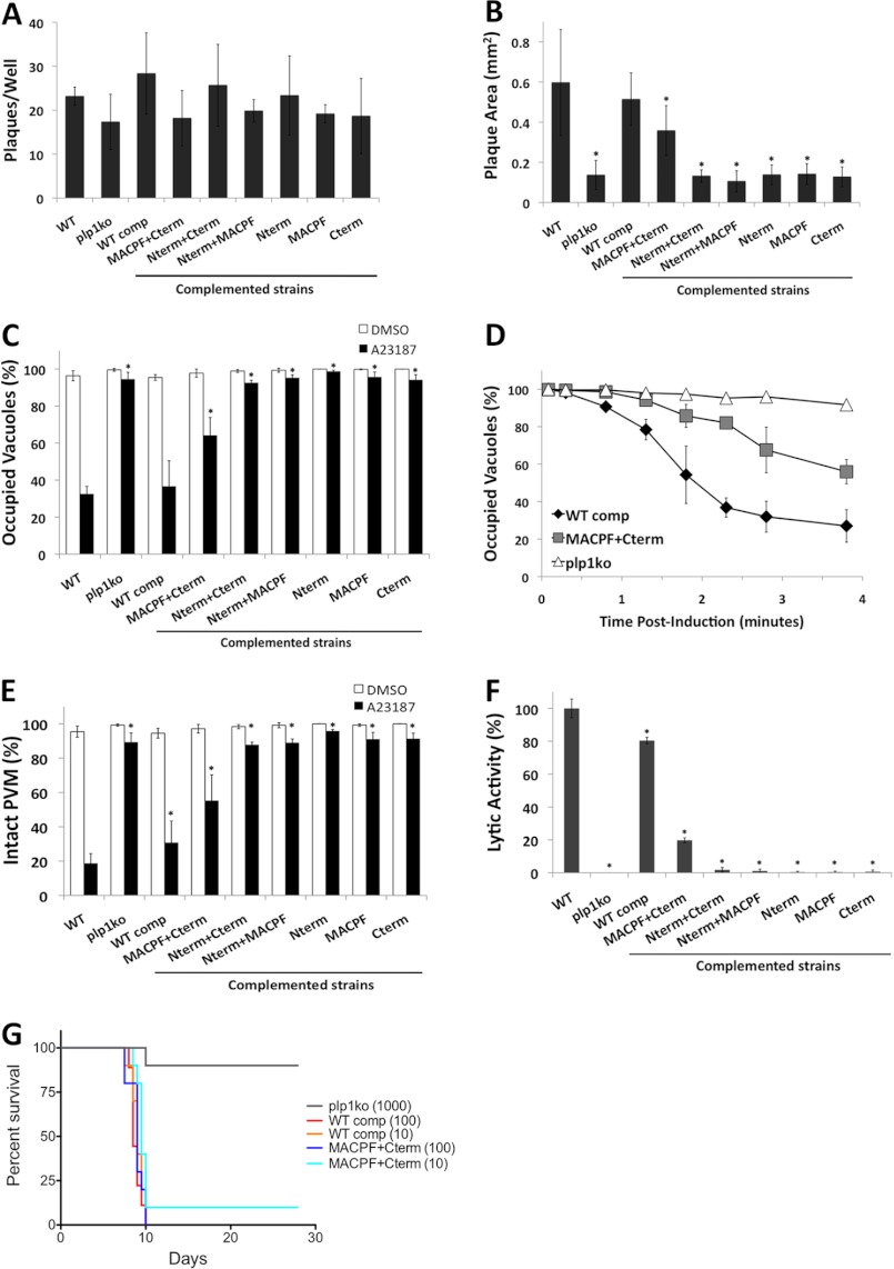
FIGURE 5.
Phenotypic analysis of PLP1 domain deletion strains reveals intermediate or nonfunctional complementation. A, 50 parasites per well were allowed to form plaques for 7 days. B, plaque area was measured by determining the average diameter in two dimensions and approximating a circle. C, egress was tested following ionophore (A23187) treatment by immunostaining vacuoles for dense granule protein 7 (GRA7) and parasites for surface antigen 1 (SAG1) and quantifying vacuoles containing parasites (occupied) and parasite-free vacuoles (unoccupied). D, time course of parasite egress is shown as in C with indicated times for each strain. E, PVM permeabilization was determined by treating infected cells with cytochalasin D prior to ionophore (A23187) treatment and fixation; infected cells were identified by bright field, and PVM permeabilization was indicated by release of the PV-targeted fluorescent protein dsRed into the host cytosol. F, lytic activity determined from PVM permeabilization was normalized to WT parasites. Graphs depict the average ± S.D. (error bars) of three independent experiments. *, p < 0.05 by one-tailed Student's t test. G, mouse survival after infection. Ten Swiss-Webster mice per strain were injected intraperitoneally with the indicated numbers of parasites and monitored for morbidity and mortality.

