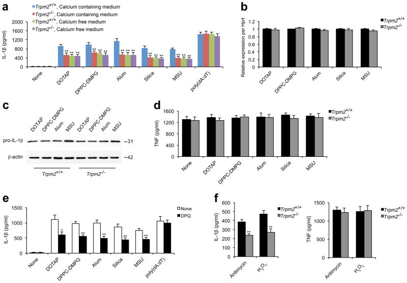
Figure 4. Ca2+ influx via TRPM2 is crucial for liposome- or crystal-induced IL-1β secretion.
ELISA for IL-1β (a) or TNF (d) from the supernatants of LPS-primed wild-type (Trpm2+/+) or Trpm2−/− BMDMs that were stimulated with indicated liposomes (30 μg/ml), crystals (alum, 250 μg/ml; silica and MSU crystals, 200 μg/ml) or poly(dA:dT) (2 μg/ml) for 6 h in calcium-containing or calcium-free medium. (b) The levels of pro-IL-1β mRNA were quantified by real-time RT-PCR in LPS-primed Trpm2+/+ or Trpm2−/− BMDMs after stimulation with indicated liposomes and crystals as in a. The gene expression data are presented as expression relative to HPRT1, and the relative gene expression levels in wild-type macrophages were designated as 1. (c) Immunoblots for pro-IL-1β and β-actin in the cell lysates from LPS-primed Trpm2+/+ or Trpm2−/− BMDMs after stimulation with liposomes or crystals as in a. (e) IL-1β from the supernatants of LPS-primed wild-type BMDMs that were pretreated with DPQ (200 μM) followed by inflammasome agonist stimulation as in a. (f) IL-1β (left) or TNF (right) from the supernatants of LPS-primed wild-type BMDMs that were stimulated with antimycin A (20 μg/ml) or H2O2 (10 mM) for 6 h. Experiments described in b–f were performed in calcium-containing medium. All data are representative of at least three independent experiments, and are shown as mean ± s.d. in a, b, and d–f. *, p < 0.05, and **, p<0.01 versus controls. Statistical significance was determined by the standard two-tailed Student’s t-test.

