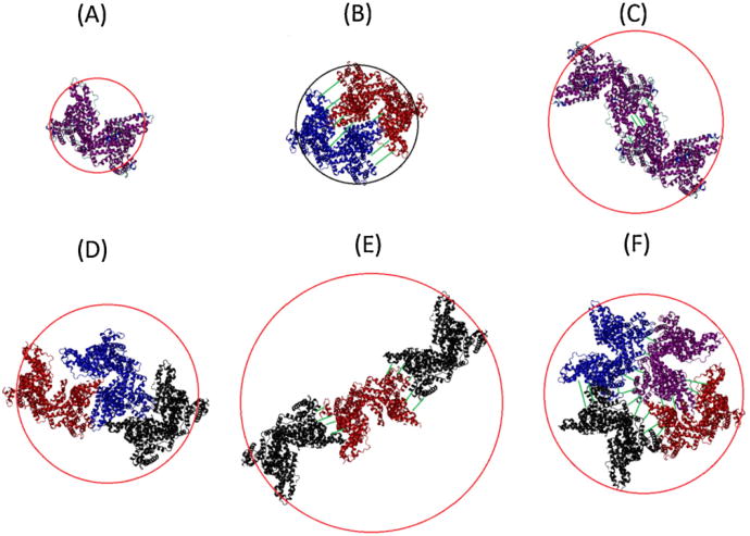
Fig. 8.
Rough visualization of possible configuration of bovine serum albumin oligomers, circled by the effective hydrodynamic diameter. The visualizations incorporate the crystal structure of HSA, which is assumed to resemble the structure of BSA. (A) monomer form, (B) dimer form fitted side to side, (C) dimer form fitted end-to-end, (D) trimers form with close association, (E) trimer formation end-to-end and (F) tightly packed tetramer form. The different colored proteins represent different subunits of the oligomer.

