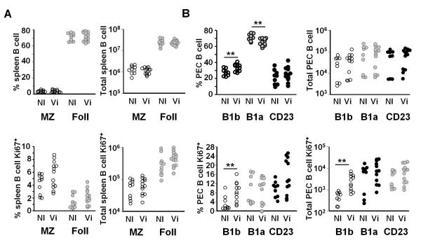
FIGURE 3.
Vi antigen selectively induces peritoneal B1b cells to proliferate. Non-immunized (NI) TCRβδ−/− mice or TCRβδ−/− mice were immunized with 10 μg Vi antigen (Vi) i.p. and 4 days later the splenic and peritoneal B cell responses were assessed by flow cytometry. A, The percentage and number of all B cells in splenic B cell subsets (top). Marginal zone B cells, MZ, were identified as IgM+CD19+B220+CD21+CD23low/− and follicular B cells as IgM+CD19+B220+CD23+CD21low B cells, Foll = follicular. The bottom graphs show the numbers and proportion of these subsets that are Ki67+. B, The number and proportion of peritoneal B1 and recirculating B cells, from the same TCRβδ−/− mice as A above, that were in the B1a, B1b and CD23 subset. All B1 cells were identified as IgM+CD19+CD21−CD23− B220+ cells and B1a cells by coexpression of CD5. CD23 cells were IgM+ CD19+ CD23+ CD21−. The bottom graphs show the number and proportion of peritoneal B1 and recirculating B cells that express Ki67. For gating protocols see Supp Fig 1 and methods. NS = not significant, ** = p ≤ 0.01 as assessed by two-tailed Mann Whitney U test. Pooled data from 3 experiments.

