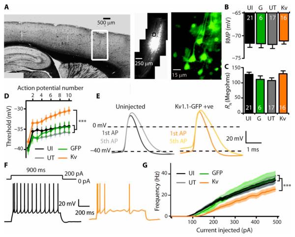
Fig. 4.
In vivo injection of Kv1.1 lentivirus attenuates excitability of adapting layer 5 pyramidal neurons. (A) GFP expression in a restricted area of motor cortex after injection of Kv1.1-GFP lentivirus. (B and C) Subthreshold properties in neurons from uninjected animals (UI) and animals injected with GFP only lentivirus (G), and in GFP-negative untransduced (UT) and neighboring GFP-positive (Kv) neurons from Kv1.1-GFP–injected animals [resting membrane potential (RMP) (B) and resting input resistance (RN) (C)]. (D to G) Neuronal excitability in neurons overexpressing Kv1.1. (D) Action potential threshold in a train of spikes (P < 0.0005 for an effect of group, linear mixed model analysis). (E) First and fifth action potentials showing depolarized threshold but no difference in accommodation (spike amplitude, rise time, decay, and width at half-maximum). (F) Representative traces from neighboring untransduced (black) and Kv1.1-overexpressing (orange) neurons in response to +200-pA current injection. (G) Frequency-current relationship showing a significant decrease in firing rate in neurons overexpressing Kv1.1 (P < 0.0001 for difference between groups, log-inear mixed model). All recordings at 36 ± 1°C, 7 to 20 days after virus injections. ***P < 0.001.

