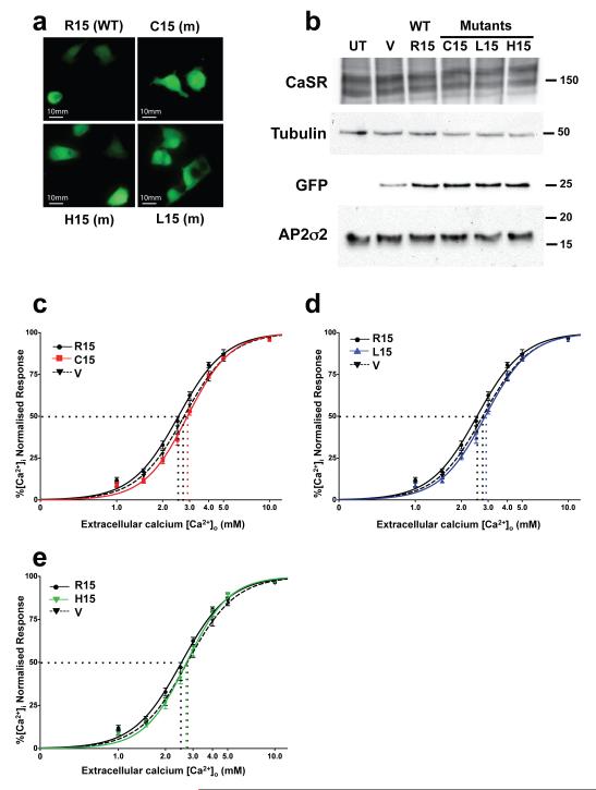
Figure 3. AP2S1 Arg15 mutants increase EC50 of CaSR-expressing cells.
HEK293 cells stably transfected with CaSR were transiently transfected with wild-type (WT) or mutant (m) AP2S1-pBI-CMV2-GFP expression vector, or the empty vector pBI-CMV2-GFP. a, Fluorescence microscopy confirmed GFP expression and successful transfection19,24. b, Western blot analysis, using anti-CaSR, anti-tubulin, anti-GFP, and anti-AP2σ2 antibodies19,24-26. HEK293 cells endogenously express AP2σ2 (Supplementary Fig.2), and co-expression of mutant AP2σ2 mimicked the situation occurring in FHH3 patients (Supplementary Fig. 1). Transfection with WT or m AP2S1-pBI-CMV2-GFP vector resulted in a significantly greater expression of AP2σ2 by 1.5 to 2.3-fold (p<0.05) when normalised for tubulin expression and compared to transfection with empty vector (data not shown). UT-untransfected cells and V-empty vector. c-e, [Ca2+]i responses (mean±SEM, n=12) to changes in [Ca2+]o of cells transfected with AP2S1 WT, mutant (m) or vector (V) alone; the vector expressed GFP but not AP2σ2, whereas the WT and mutant constructs expressed GFP and either WT or mutant AP2σ2, respectively. c, Cys15 (C15), d, Leu15 (L15), and e, His15 (H15) constructs. EC50 values (dotted lines).

