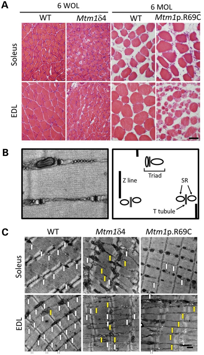
Figure 3.

Light microscopic pathology in myotubularin-deficient mice is dependent on mouse strain and the muscle examined. (A) Hematoxylin and eosin staining of EDL and soleus muscles from Mtm1δ4, Mtm1 p.R69C or age-matched WT littermate mice display variable levels of myofiber smallness and centrally nucleated fibers. Note that differences in fiber size when comparing the two WT populations is due to the normal growth between 6 weeks of life (WOL) and 6 months of life (MOL). (B) To evaluate sarcotubular organization at the ultrastructural level, the number of triads, T-tubules and L-tubules were quantified from longitudinal sections of EDL and soleus muscles from WT and Mtm1δ4 animals at six WOL and WT and Mtm1 p.R69C animals at six MOL. A picture and diagram of the normal sarcotubular architecture from a 6-week-old WT mouse is shown. (C) Representative images (×9300 magnification) of longitudinal sections of 6-week-old WT and Mtm1δ4 mice, which allow the quantification of T-tubules (white arrows) and L-tubules (yellow arrows). Representative images from a 6-month-old Mtm1 p.R69C mouse are also shown for comparison. Bar = 50 μm in (A) and 500 nm in (C).
