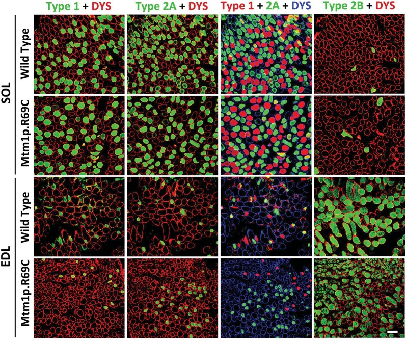
Figure 4.
Immunohistochemical evaluation of fiber types in Mtm1p.R69C EDL and soleus muscles. Immunohistochemistry for dystrophin (red or blue) and either type 1, type 2A or 2B (green) myosin reveals the fiber-type populations present within the EDL and soleus muscles of Mtm1p.R69C and age-matched WT mice. Fiber-type proportions were similar in 6-week-old Mtm1δ4 and WT mice. A pseudocolored overlay from adjacent muscle sections stained with type 1 and 2A myosin is provided to depict the total population of oxidative fibers in these muscles. Similar distributions of oxidative and glycolytic fibers were found in WT and Mtm1δ4 EDL and soleus muscles at 6 weeks of life, as described in Table 2. Bar = 100 μm.

