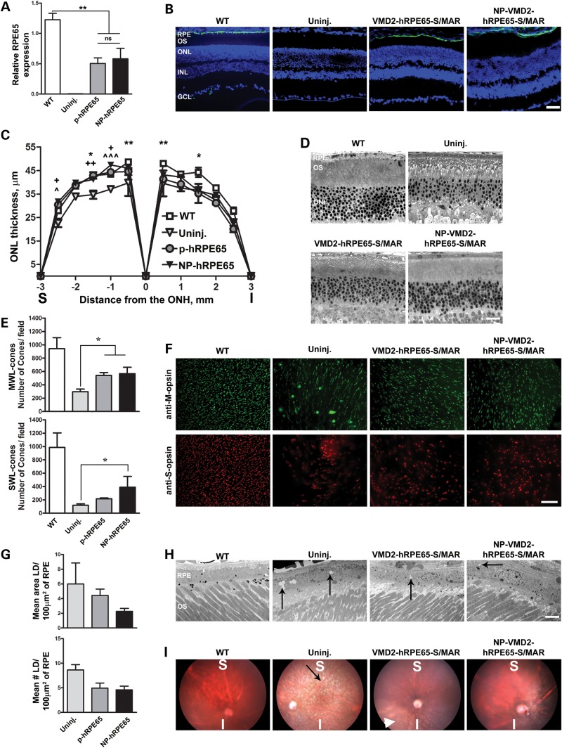
Figure 3.
S/MAR-containing hRPE65 vectors mediate improvement in structural phenotypes in the rpe65−/− model. (A) qRT-PCR was conducted at PI-180 on whole eyes from rpe65−/− animals injected at P16 with S/MAR-containing hRPE65 vectors using primers that amplify from both mouse and human RPE65. **P < 0.01 by one-way ANOVA with Bonferroni's post hoc comparison. (B) IHC showing the distribution of RPE65 (green) at PI-180 in rpe65−/− mice using Abcam antibody 13826 against human and mouse RPE65. (C) ONL thickness was measured at PI-180 at increasing distances from the optic nerve head (ONH). Asterisk indicates significant differences between WT and uninjected, plus symbol indicates significant differences between NP and uninjected, arrow head symbol indicates significant differences between naked and uninjected. One symbol, P < 0.05, two symbols, P < 0.01, three symbols P < 0.001 by two-way ANOVA with Bonferroni's post hoc tests. n = 3–5 eyes/group. (D) Representative images used for quantification in (C). (E and F) PI-180 retinal whole mounts were labeled with antibodies against MWL cone opsin (upper panels F, green) and SWL cone opsin (lower panels F, red). Cones were counted in 16 images (four/quadrant) from each eye using ImageJ. Values were averaged to give mean cone counts/field for the whole eye. n = 4 eyes/group. Shown in (E) are the mean ± SEM. *P < 0.05 in one-way ANOVA with Bonferroni's post hoc test. (G and H) RPE lipid droplets (arrows, H) were counted and their area measured in 10–15 EM images/eye (4 eyes/group) at PI-180. Representative EMs shown in (H). The mean area occupied by the lipid droplets/100 µm2 of RPE is shown in the top panel of (G), while the mean number of lipid droplets/100 µm2 of RPE is shown in the bottom panel of (G) (±SEM). (I) Fundus images were obtained at PI-180 showing improvement in lipid droplet accumulation at the region of injection. Black arrow shows white spicules indicative of retinyl ester accumulation; white arrowhead shows the same phenotype in the inferior of a VMD2-hRPE65-S/MAR-treated eye. Scale bars: (B) 40 µm; (D) 20 µm; (F) 50 µm; (H) 200 nm. RPE, retinal pigment epithelium; OS, outer segment; IS, inner segment; ONL, outer nuclear layer; INL, inner nuclear layer; GCL, ganglion cell layer.

