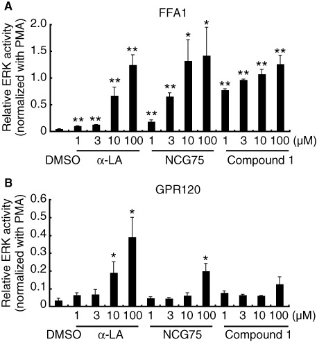
Figure 2.

Effect of compounds on ERK activity in (A) T-REx FFA1 and (B) Flp-in GPR120 cells. Cells were serum-starved for 20 h and then treated with various compounds at 1–100 µM. Cell lysates were analyzed by immunoblotting using anti-phospho- and anti-total-kinase antibodies. The amount of phosphorylated ERK was normalized to the amount of total ERK. The data were then presented as -fold difference relative to the amount of ERK phosphorylation that was obtained in the presence of phorbol 12-myristate 13-acetate (PMA). Results represent means ± SEM of three independent experiments. *P < 0.05, **P < 0.01, significantly different from control (DMSO).
