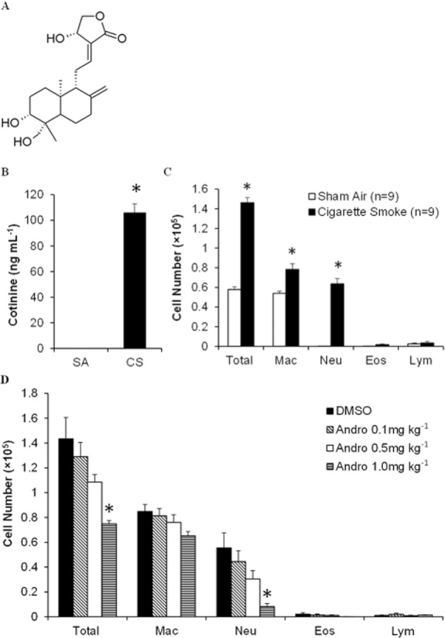
Figure 1.

Effects of andrographolide on cigarette smoke-induced inflammatory cell recruitment. (A) The chemical structure of andrographolide. (B) Blood was collected immediately after 1 h 4% cigarette smoke exposure, and plasma cotinine levels were measured using elisa (n = 6). (C) Inflammatory cell counts in BAL fluid obtained from mice 24 h after the last sham air (n = 9 mice per group) or cigarette smoke (n = 9 mice per group) exposure. (D) Andrographolide dose-dependently reduced cigarette smoke-induced inflammatory cell counts in BAL fluid from mice 24 h after the last cigarette smoke challenge (DMSO, n = 6; 0.1 mg·kg−1, n = 6; 0.5 mg·kg−1, n = 6; and 1 mg·kg−1, n = 8 mice per group). Differential cell counts were performed on a minimum of 500 cells to identify eosinophil (Eos), macrophage (Mac), neutrophil (Neu) and lymphocyte (Lym). Values shown are the mean ± SEM. *Significant difference from DMSO, P < 0.05.
