
Figure 3.
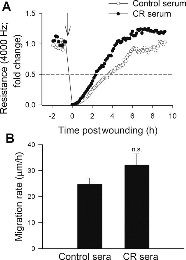
Effect of treatment with sera collected from caloric-restricted (CR) Macaca mulatta on migration capacity of human coronary arterial endothelial cells (CAECs). Vascular endothelial growth factor (VEGF; 100ng/mL)-stimulated cell migration was monitored by electric cell–substrate impedance sensing (ECIS) technology in a wound-healing assay (see Methods section). (A) Representative figure showing the time course of resistance recovery after wounding (arrow indicates the time, when an electric pulse of 5 mA for 20 seconds at 100kHz was applied; 100% represents prewounding levels of resistance measured at 4000 Hz). Resistance (at 4000 Hz) was monitored for every 160 seconds. Time to reach 50% resistance recovery (corresponding to 50% confluence on the active electrode) was determined for CAECs treated with control sera or CR sera, and this parameter and the known physical dimensions of the electrode were used to calculate the migration rate (expressed as µm/h). (B) The summary data for migration rate in CAECs treated with control sera or CR sera are depicted. Data are means ± SEM (n = 5 in each group).