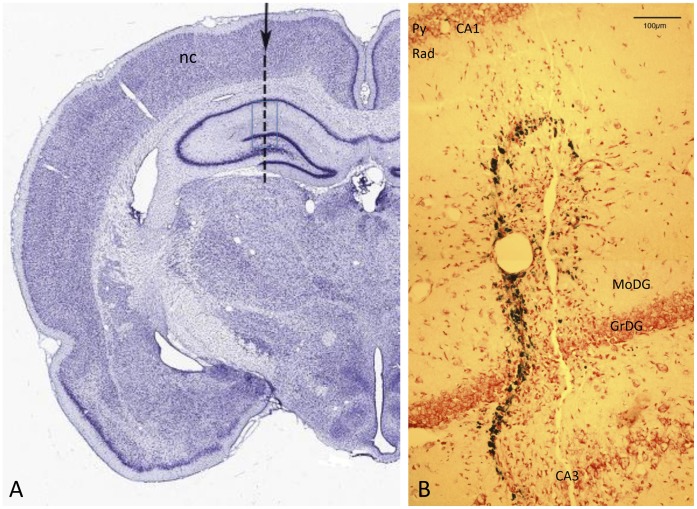
Figure 1. The needlestick lesion.
A: Coronal section of rat brain, ∼3 mm anterior to bregma, stained with Thionine [74]. The arrow and dotted line show the targeted position of the needle track. The track traverses neocortex (nc) and hippocampus, from CA1 across the dentate gyrus and CA3. The blue rectangle shows the area imaged at higher power, from an experimental animal, in B. B: Needle track crossing layers of the hippocampus, in the region marked by the rectangle in A. The track is evident as a string of Prussian-blue labelled deposits, close to a linear break in the neuropil. Neutral red counterstain. Py – pyramidal layer of CA1; Rad - stratum radiatum of CA1; MoDG – molecular layer of the dentate gyrus; GrDG – granular layer of the dentate gyrus.

