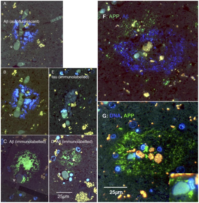
Figure 10. Cellular organisation of human plaques, seen in serial, thin (0.1 µm) sections.
A–E are sections of one plaque, F and G of another. A: The blue fluorescence is autofluorescence from amyloid in the plaque. It surrounds a branch point of a capillary-size vessel (v). B: 0.8 µm away, the amyloid autofluorescence is still prominent. C: 1.0 µm from A (and 0.2 µm from B) the plaque is labelled green with the 4G8 antibody. D: 9 µm from A (in the same direction), the plaque remains clearly labelled with the 4G8 antibody. E: 4 µm from A, the same vessel (v) is apparent; small elements of tau labelling (green) are shown, in the area of the plaque. F: Blue autofluorescence shows amyloid forming a shell surrounding red and yellow autofluorescent bodies. Labelling for APP (green) shows punctate APP+ elements forming an incomplete shell outside the amyloid shell. G: The blue fluorescence in this panel is bisbenzimide labelling for nuclear DNA. 4G8 labelling (green) shows Aβ forming a shell around a cellular core, which includes the vessel, some nuclei and autofluorescent material. The inset shows a vessel at the centre of the plaque, at higher power.

