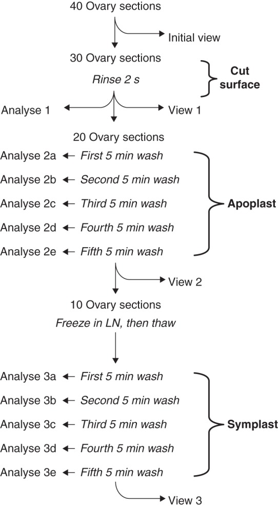
Fig. 2.

Protocol for locating glucose or CF in the apoplast and symplast of live sections of maize ovaries. The central column shows the rinse and wash-outs of the sections while the left side shows when the wash water was analysed (Analyze 1–3e), and the right side shows when sections were removed for imaging (Initial view and Views 1–3). The rinse removed surface glucose or CF (Cut surface), and the next five washes removed glucose or CF from the apoplast (Apoplast). After freezing in liquid nitrogen (LN) and thawing to break cell membranes, the final five washes removed glucose or CF from the symplast (Symplast). At the end of the protocol, glucose or CF was extracted from the last ten sections (View 3) for a final analysis. The protocol was identical for glucose or CF but used different plants.
