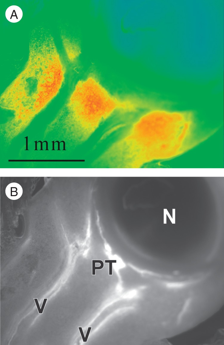
Fig. 3.

Apoplast glucose in a live maize ovary on the day of pollination, after a 2-s rinse to remove glucose from cut cells in the section. Because the enzymes for the glucose assay could not enter the live cells, only apoplast glucose is visible. (A) Glucose (red) is abundant in the upper tissues of the pedicel. (B) UV autofluorescence of the vascular system (V) and phloem termini (PT) of the section in (A) shows no vascular bundles enter the nucellus (N).
