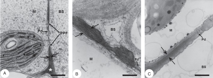
Fig. 3.

Transmission electron micrographs of leaf blade cross-sections showing the bundle sheath–mesophyll wall interface in Trianthema portulacastrum, Trianthema sheilae and Zaleya pentandra. (A) Trianthema portulacastrum wall interface with pitting and plasmodesmata. (B) Trianthema sheilae wall interface with pitting and plasmodesmata showing pronounced wall thickening in the common wall between mesophyll and bundle sheath. (C) Zaleya pentandra wall interface with pitting and plasmodesmata. Abbreviations: BS, bundle sheath; M, mesophyll; Pd, plasmodesmata; PPF, primary pit field. Scale bars: (A) = 0·5 µm; (B) = 0·25 µm; (C) = 0·4 µm.
