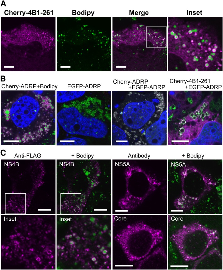
Fig. 1.
Full-length NS4B localizes to LDs. (A) Localization of Cherry-4B1–261. Oc cells were transfected with Cherry-4B1–261 for 24 h followed by staining with Bodipy493/503. (A, inset) magnified image. Scale bars, 10 μm. (B) Colocalization of Cherry-4B1–262 with ADRP. From left, Cherry-ADRP, EGFP-ADRP, Cherry-ADRP with EGFP-ADRP (cotransfection), and Cherry-4B1–261 with EGFP-ADRP (cotransfection) was transfected into Oc cells. Scale bars, 10 μm. (C) Localization of NS4B, NS5A, and Core protein. Oc cells were transfected with pcDNA-4B1–261-FLAG (left), pcDNA-5A-myc (right, top), or pcDNA-CORE (right, bottom) for 24 h. After fixation, the antigens were visualized with anti-FLAG (NS4B), anti-Myc (NS5A), or anti-Core antibodies and Alexa-Fluor-dye-labeled secondary antibodies. Scale bars, 10 μm. Inset, magnified image.

